Research

Sort research by Biological Questions  -   Sort research by Technical Approaches


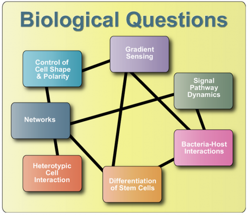
Signaling Pathway Dynamics


Signal transduction pathways, as the name implies, are often thought of as chains of molecules that simply relay external signals to end effectors.  However, this is a misnomer because signaling pathways execute other important functions (e.g. signal amplification, signal processing, etc.) and can implement critical systems features (e.g. robustness, sensitivity, memory, etc.).  Understanding such properties is crucial to understanding the biology of signaling pathways and they are best studied through signal pathway dynamics.  We are interested in how dynamic input and output, such as oscillatory, pulsatile, and switch-like behavior, relates to topology, function, and regulation especially in the NF-kappaB, MAPK, and calcineurin pathways.  Experimentally, we probe these pathways through a combination of live cell imaging, immunocytochemistry, Western blots, real time PCR, and other techniques.  The resulting data is analyzed in the context of computational models of the pathway.  In parallel, we are developing mathematical theory that can be used to evaluate pathway dynamics and system properties.
 

Gradient Sensing


Gradient sensing by cells involves the ability to sense extremely small spatiotemporal differences in chemical or physical cues in a precise manner. Cells respond to external chemical gradients by polarizing and migrating toward chemoattractants or away from chemorepellants (chemotaxis) or undergoing directed morphogenesis (chemotropism). These phenomena are crucial for proper functioning of single-cell organisms, such as bacteria, amoebae and yeast; as well as multi-cellular systems as complex as the immune and nervous systems. Chemotaxis is also important in wound healing and tumor metastasis. In addition, the migratory behavior as well as morphogenetic growth of most of the cells in the body depends on the properties of the extracellular matrix. Mechanotransduction induced cues are important for phenomena like durotaxis (cell motility or growth up or down a rigidity gradient) and haptotaxis (up or down a gradient of cellular adhesion sites).
Traditionally, gradient sensing studies have been affected by lack of appropriate control of spatial (the actual shape of the gradient) or temporal gradients (gradient stability). Our lab has been developing a range of novel microfluidic device designs, which are tailor-made to allow accurately controlled gradient sensing studies for different cell-types. In addition, these studies are complemented by molecular biology approaches and mathematical modeling of the signal transduction pathways to understand the underlying basis of gradient sensing in these cells.
We study a diverse range of gradient sensing systems, ranging from pheromone sensing in yeast and axon guidance in embryonic Xenopus spinal neurons to the mammalian gradient sensing pathways involved in angiogenesis, tumor metastasis, haptotaxis and durotaxis.
 

Bacteria-Host Interactions


We attempt to deepen the understanding of how bacteria interact with the host system in the context of infection. Bacterial pathogens make use of several strategies such as adhesions to host cells, cellular invasion followed by intracellular proliferation, adaptation, or persistence. To investigate these exquisite strategies of bacterial pathogens, we implement the precise control of chemical and mechanical cues on the microfluidic design for bacteria culture and bacteria-host co-culture environment. High throughput approaches using this microfluidic platform allow us to overcome the drawbacks of the traditional drug screening and experimentation with cells in suspension or on gel plates. Using quantitatively characterized cellular and molecular events based on the experiment approaches, we take into account various interactions, including those relying on various feedback loops and associated adaptive behavior, between bacterial pathogens and host systems.
 

Differentiation of Stem Cells


The process by which stem cells differentiate into various tissues is largely unknown due to the enormous complexity of signals, both chemical and mechanical.  To add to this complexity, these signals change quickly overtime and are finely controlled by dosage.  Current methods in cell culture conditions do not easily afford such fine control of the developing stem cells which results in experiments that are both time-consuming and expensive.  To overcome these difficulties, our group is exploring the use of microfluidic devices and nanotechnology to finely control the chemical and physical environment of embryonic and mesenchymal stem cells.   Currently these methods are being applied to understand the effect of various soluble factors such as Leukemia Inhibitory Factor (LIF) and Bone Morphogenic Protein 4 (BMP4) on self-renewing cells.   Additionally, this group is exploring the soluble factors that affect the choice of stem cells to differentiate into the various germ layers, especially neuroectoderm or mesoderm.  From these germ layers, cells such as neurons or cardiomyocytes can be derived which could have potentially dramatic clinical applications.
 

Control of Cell Shape & Polarity


Cells have the inherent ability to dynamically respond to changes in the extracellular environment.  They change shapes to better adapt to environmental cues so they can carry out their tasks with efficiency.  These cell shape changes are the result of dynamic reorganization of cytoskeleton proteins such as actin and microtubule regulated by a number of signaling molecules embedded in them.  We are studying the molecular basis of cell shape changes in response to extracellular stimuli (chemical or mechanical in nature) in different model systems such as baker’s yeast and mammalian cells.  In our experimental approaches we are using a variety of tools to study more efficiently the underlying cell biology that leads to cell shape change.  For example, in addition to standard biochemical, molecular and microscopy techniques we are using microfluidics and bioengineering tools to mimic in-vivo extracellular stimuli in in-vitro cell culture system.  Fluorescent protein tracking and detection is widely employed as reporter for gene expression in our studies.  We also develop methods for quantitative examination of dynamic fluorescent protein expression and localization in living cells.  For understanding gene function we are employing PCR-mediated gene disruptions to create either null or mutated alleles.
 

Heterotypic Cell Interaction


Cell behavior and functions are affected by its environment, which includes neighboring cells. This effect can be done by contact or by secreting soluble molecules. We investigate angiogenesis and tumor metastasis by studying interaction between tumor cells and endothelial cells for a wide range of scales: from single cell, to a group of cells to colonies. We are also interested in interactions between neuron and muscle cells, stem cell and differentiated cells, etc.
 

Networks


Biological networks are graphical representations of complex systems and processes, such as gene regulation, metabolism, or signal transduction. Large-scale topological analysis of these networks has revealed that they are not randomly organized. The intricate relations between their structure, function and dynamics need to be further investigated.

We study the dependence and interplay between network topology and the dynamics of the biological processes within a cell. We investigate ways in which network topology may have evolved to optimize the robustness of network dynamics. In addition, we investigate the notion of a close interplay between different kinds of biological networks: Specifically, we analyze how they interact as the cell receives and processes information about changes in its environment, and responds to them in optimal ways. To this end, we extensively study both the large-scale topology of biological networks and the function and dynamics of smaller topological motifs within them.

In our studies, we apply mathematical models, sophisticated simulation and visualization techniques, as well as our knowledge of the biochemical principles underlying network function. Our experimental facilities and resources permit us to change a cell’s environment in a controlled way, thus targeting different regions within the underlying biological network. By observing both single cells and in cell colonies, we can analyze diverse aspects of network dynamics.


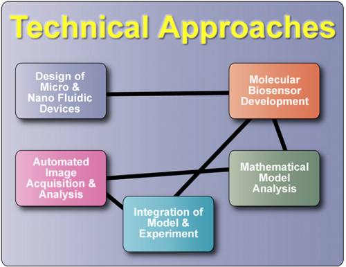
Design of Micro & Nanofluidic Devices


Intracellular signal transduction involves a variety of molecular-level events occurring dynamically in space and time. To understand the signaling processes that, when integrated, generate specific cellular response, one often needs to perform multiple experimental perturbations of signaling network.  The low throughput of traditional manual experimental technique is the limiting bottleneck in the collection of data necessary for constructing quantitative models of signaling transduction.  Commercial “robotics” is too expensive and space consuming for typical laboratories, and often only capable of performing a single task.

 
We therefore set out to design PDMS based microfluidic devices and develop the accompanying protocols for employing these devices to culture and extract data from a variety of biological model systems:  primary neuron cells, tumor cells, stem cell, immortalized cell lines, bacteria, yeast, and Dictyostelium.  The small scale and precise chemical delivery capability conferred by microfluidics enabled us to perform experiments with complex perturbation over the microenvironment the cells experience and thus revealing novel and biologically significant phenomenon.  We then proceed to probe the underlying fundamental mechanism by using our patented automation devices that allows rapid and inexpensive testing of a large number of stimulation conditions, resulting in data that facilitate the development of computational models for elucidating the complex dynamics of signal transduction.
 
Through close collaboration with Kahp-Yang Suh at Seoul National University, we have started to probe model systems at the nano scale with nanostructured polymeric substrate.  In addition, we have interfaced nanotopology with microfludic structures, creating cutting edge tools for investigating novel mechantransduction phenomenon on the nanometer scale.
Our research laboratory is situated inside the same building that houses a sophisticated microfabrication laboratory (see Facilities for further details).  Thus our turn around time from designing a new microfluidic chip to finished product is typically 1 work day.  We also possess an array of sophisticated microscopy systems that are coupled to our custom microfluidic setup (see Automated Image Acquisition & Analysis), along with cell culture laminar flow hoods for preparing our in-chip experiment.  Our uniquely integrated environment enables us to rapidly create microfluidic tools that closely fulfill the needs of the mainstream biologists.
 

Automated Image Acquisition & Analysis


Our lab has several fully automated epifluorescent deconvolution microscopes, ranging from x-y-z stage, filter and multi-camera control. The scopes are driven through 3I’s Slidebook and custom software. When coupled to our robust microfluidic platform, a single experiment can generate thousands of images with exquisite spatial-temporal detail. Following data acquisition, we seek to determine quantitative and significant relationships from our image data, measuring protein expression, cell morphology, and motility patterns. The lab is actively implementing and investigating a large variety of imaging algorithms, especially PDE based segmentation involving parametric and geometric active contours.
 

Integration of Model & Experiment


In our lab, experimental and mathematical approaches are integrated to explore the signaling pathways within a single cell and communications among cells. Microfluidic systems, combined with other techniques (eg. real time PCR, live cell imaging, Western blots, and immunocytochemistry), are utilized to better define the environment of cell culture and to characterize the molecular events more quantitatively in both spatial and temporal dimensions. Computational models are developed based on experimental results, providing better understanding of the underlying mechanisms and making predictions to be validated by further experiments. With the power of integrated experimentation and simulation in mathematical models, we have successfully developed insight into the roles of MAPK cascades in pheromone sensing of yeast and NF-kappaB pathway in inflammation. We expect this approach will also be fruitful in other areas we are studying, such as angiogenesis, axon guidance, tumor metastasis and cell morphogenesis.


Mathematical Model Analysis


We use a wide variety of different mathematical approaches to address many aspects of the biological questions we are currently investigating.  Systems of ordinary differential equations (ODEs) are used to predict the behavior of complex intracellular signaling pathways and validated through both conventional and novel experimental approaches.  For biological questions that are spatially dependent, such as chemotaxis and axonal guidance, we use both finite element and finite volume approaches to determine the significance of the polarization of signaling molecules. Depending on the question at hand we have also utilized a finer scale analysis involving Monte Carlo simulations tracking individual interacting molecules or agent based modeling aimed at describing multiple interacting cells within a population.  In addition, we also use network analysis to model regulation of gene transcription and validate results using molecular biosensors.

Molecular Biosensor Development


Analysis of intercellular signaling events requires development of novel probes for efficient tracking of localization and activity of various molecular players. We are using modern molecular biology tools to create a library of molecular biosensor that would allow us to image multiple signaling events, on a large scale (hundreds to thousands of cells) simultaneously. When coupled to our micro- and nano-scale device technology, utilization of the biosensors results in precisely controlled datasets providing a wealth of information on signaling events expressed in the form of imaging information with exquisite temporal and spatial resolution.